Discover Geographic Atrophy

Geographic Atrophy (GA) is an advanced form of age-related macular degeneration (AMD), a leading cause of significant vision loss worldwide and in Canada.1-3
Explore new territory in GA
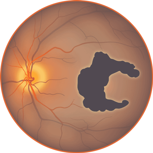
GA progression is relentless and irreversible4-7
While lesion growth may appear to proceed slowly, GA is progressive and the associated vision loss is irreversible.4-7
GA affects

There is currently
no treatment for GA in Canada
Excessive activation of the complement system
may lead to lesion progression and loss of vision4,8-10
Learn about the role of the complement system in GA.

This is Geographic Atrophy, or GA, when damage to the retina is irreversible. To uncover what’s behind the destruction, let’s look below the surface.
In this advanced form of dry age-related macular degeneration, atrophic lesions form on the macula, causing loss of functional vision as they spread. This group of proteins is called the complement system. Normally, they defend against pathogens and remove abnormal cells, yet in Geographic Atrophy, or GA, too much complement activity can be detrimental.
This central protein, C3, plays a key role in the excessive complement activity. Complement activation causes C3 to cleave, splitting it apart into the active fragments, C3a and C3b. Over by this photoreceptor, a group of C3a’s have begun to accumulate. Too much C3a can trigger chronic inflammation of the macula that can contribute to cell death.
Here, a group of C3b’s are accumulating on cell surfaces, tagging them for phagocytosis by immune cells. But the journey is far from over for C3b, which also has significant effects on the complement cascade, causing downstream components to work together and form something new, the membrane attack complex, that latches onto and forms an opening in the cellular membranes of retinal pigment epithelial cells, or RPE, as well as photoreceptors.
C3 is a central protein in the complement system, but when there is excessive complement activation, C3 plays a key role in inflammation, phagocytosis, and cell membrane disruption.
All of which are thought to ultimately lead to retinal cell death.
Hear from
your peers

Why it is critical to
recognize GA at first sight
Understand how to identify patients with GA and monitor progression.


Apellis is a global biopharmaceutical company that leverages courageous science and creativity. We are committed to addressing the unmet needs of patients and eye care professionals worldwide.
Sign up to stay
connected
Receive updates about GA from Apellis.

References:
- Gehrs KM, Anderson DH, Johnson LV, Hageman GS. Age-related macular degeneration—emerging pathogenetic and therapeutic concepts. Ann Med. 2006;38(7):450-471. doi:10.1080/07853890600946724.
- Fleckenstein M, Mitchell P, Freund B, et al. The progression of geographic atrophy secondary to age-related macular degeneration. Ophthalmology. 2018;125(3):369-390. doi:10.1016/j.ophtha.08.038.
- Noble J, Chaudhary V. Five things to know about age-related macular degeneration. CMAJ. 2010;182(16):1759.
- Boyer DS, Schmidt-Erfurth U, van Lookeren Campagne M, et al. The pathophysiology of geographic atrophy secondary to age-related macular degeneration and the complement pathway as a therapeutic target. Retina. 2017;37(5):819-835. doi:10.1097/iae.0000000000001392.
- Lindblad AS, Lloyd PC, Clemons TE, et al. Age-Related Eye Disease Study Research Group. Change in area of geographic atrophy in the age-related eye disease study: AREDS report number 26. Arch Ophthalmol. 2009;127(9):1168-1174. doi:10.1001/archophthalmol.2009.198.
- Holz FG, Strauss EC, Schmitz-Valckenberg S, van Lookeren Campagne M. Geographic atrophy: clinical features and potential therapeutic approaches. Ophthalmology. 2014;121(5):1079-1091. doi:10.1016/j.ophtha.2013.11.023.
- Sunness JS, Margalit E, Srikumaran D, et al. The long-term natural history of geographic atrophy from age-related macular degeneration: enlargement of atrophy and implications for interventional clinical trials. Ophthalmology. 2007;114(2):271-277. doi:10.1016/j.ophtha.2006.09.016.
- Katschke KJ Jr, Xi H, Cox C, et al. Classical and alternative complement activation on photoreceptor outer segments drives monocyte-dependent retinal atrophy. Sci Rep. 2018;8(1):7348. doi:10.1038/s41598-018-25557-8.
- Park DH, Connor KM, Lambris JD. The challenges and promise of complement therapeutics for ocular diseases. Front Immunol. 2019;10:1007. doi:10.3389/fimmu.2019.01007.
- Yates JRW, Sepp T, Matharu BK, et al. Genetic Factors in AMD Study Group. Complement C3 variant and the risk of age-related macular degeneration. N Engl J Med. 2007;357(6):553-561. doi:10.1056/NEJMoa072618.